40 external structure of the heart with labels
Surface projections of the heart: Borders and landmarks - Kenhub The right margin of the heart consists of two lateral convex arches separated by an obtuse angle. In adults, the lower arch belongs to the right atrium, while the upper arch represents the ascending aorta. However, in children, the upper arch is actually flat and is formed by the superior vena cava. Coronary arteries and cardiac veins: Anatomy and branches - Kenhub This vessel was first described back in 1928 as a large calibre vessel that travels in the walls of the atrial appendage and provides indirect and direct anastomotic pathways between the left and right coronary arteries. Once on the diaphragmatic surface, the left circumflex artery travels along the diaphragmatic atrioventricular groove.
Heart Labeling Quiz: How Much You Know About Heart Labeling? Here is a Heart labeling quiz for you. The human heart is a vital organ for every human. The more healthy your heart is, the longer the chances you have of surviving, so you better take care of it. Take the following quiz to know how much you know about your heart. Questions and Answers 1. What is #1? 2. What is #2? 3. What is #3? 4. What is #4?

External structure of the heart with labels
Diagrams, quizzes and worksheets of the heart - Kenhub Using our unlabeled heart diagrams, you can challenge yourself to identify the individual parts of the heart as indicated by the arrows and fill-in-the-blank spaces. This exercise will help you to identify your weak spots, so you'll know which heart structures you need to spend more time studying with our heart quizzes. How the Heart Works - The Heart | NHLBI, NIH The heart is an organ about the size of your fist that pumps blood through your body. It is made up of multiple layers of tissue. Your heart is at the center of your circulatory system. This system is a network of blood vessels, such as arteries, veins, and capillaries, that carries blood to and from all areas of your body. Heart anatomy: Structure, valves, coronary vessels | Kenhub The heart is shaped as a quadrangular pyramid, and orientated as if the pyramid has fallen onto one of its sides so that its base faces the posterior thoracic wall, and its apex is pointed toward the anterior thoracic wall.
External structure of the heart with labels. Anatomy Of Heart Quiz! - ProProfs Welcome to Anatomy of Heart Quiz! The heart is charged with the sole function of pumping blood throughout the body and, in so doing, ensuring nutrients can be transported with ease. It carries deoxygenated blood to the lungs, where it loads up with oxygen and unloads carbon dioxide. Test your knowledge of the heart anatomy by taking up the quiz below. All the best, and good luck! Layers of the heart: Epicardium, myocardium, endocardium - Kenhub The endocardium is the innermost layer of the heart. It lines the inner surfaces of the heart chambers, including the heart valves. The endocardium has two layers. The inner layer lines the heart chambers and is made of endothelial cells. Female Body Diagram: Parts of a Vagina, Location, Function Vagina: The vagina is a muscular canal that connects the cervix and the uterus, leading to the outside of the body. Parts of the vagina are rich in collagen and elastin, which give it the ability to expand during sexual stimulation and childbirth. Cervix: The cervix is the lower part of the uterus that separates the lower uterus and the vagina and may play a role in lubrication. The Ultimate Heart Model & Sheep Heart Practice Quiz! - ProProfs As is the case in the lab practical, each correct answer counts. So, make sure you learn from the feedback. Questions and Answers 1. 1. Name the structure- be specific. 2. 2. Name the vessel- be specific. 3. 3. Name the structure-be specific. 4. 4. Name the vessels. 5. 5. Do the vessels in #4 carry blood to or from the heart? A. To the heart B.
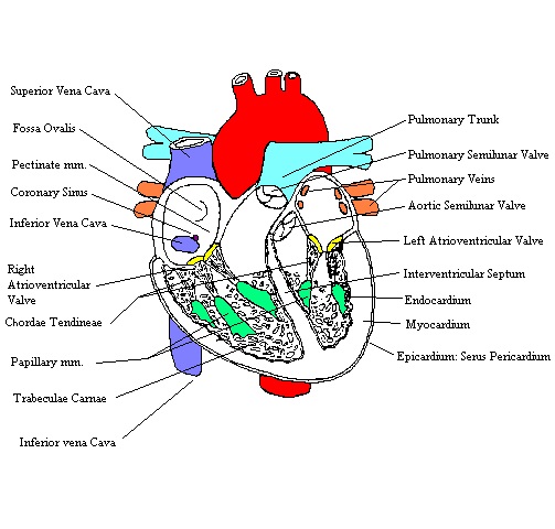
Heart: Anatomy | Concise Medical Knowledge - Lecturio The heart is a 4-chambered muscular pump made primarily of cardiac muscle tissue. The heart is divided into 4 chambers: 2 upper chambers for receiving blood from the great vessels, known as the right and left atria, and 2 stronger lower chambers, known as the right and left ventricles, which pump blood throughout the body. Body Cavities and Membranes: Labeled Diagram, Definitions The 3 meningeal layers are labeled with the stars. The outermost layer of the meninges is the dura mater, which is located beneath the skull. Below the dura mater is the arachnoid, which is the middle meningeal layer. There is a space below the arachnoid called the subarachnoid space, and this is where the CSF is located. Normal CT chest lung on axial images with labels | e-Anatomy - IMAIOS Anatomy of the chest: how to view the anatomical labels. This atlas is a comprehensive and affordable learning tool for medical students and residents and especially for radiologists and pneumologists. It provides access to CT images in the axial plane, allowing the user to learn and review the lung anatomy interactively. External Female Genital Anatomy - Study.com The female's external anatomy consists of six main structures. The labia minora and the labia majora help to protect and moisten the vulvar area. Extensions of the labia minora make up the prepuce ...
Anatomy, Blood Vessels - StatPearls - NCBI Bookshelf Structure and Function. Vessels transport nutrients to organs/tissues and to transport wastes away from organs/tissues in the blood. A primary purpose and significant role of the vasculature is its participation in oxygenating the body. Deoxygenated blood from the peripheral veins is transported back to the heart from capillaries, to venules, to veins, to the right side of the heart, and then ... Anatomical Position and Directional Terms - EZmed Medial vs Lateral. The first pair of directional terms is medial and lateral. To better understand medial and lateral, let's divide the body into right and left sections using a sagittal plane.. You might remember in the previous lecture on body planes that the sagittal plane runs vertically front to back, and it divides the body into right and left sections. Kidney Structures and Functions Explained (with Picture and Video) The glomerulus connects to a long, convoluted renal tubule which is divided into three functional parts. These consist of the loop of Henle (nephritic loop), the proximal convoluted tubule, and the distal convoluted tubule, which empties into the collecting ducts. These collecting ducts fuse together and enter the papillae of the renal medulla. Correctly Label The Following Internal Anatomy Of The Heart The aorta, or aortic arch, is the outermost layer of the heart. The left ventricle is covered with the ventricular aorta, and the pulmonary veins are located inside the aorta. The two atria, the left and right aorta, and the right aortic arch are all external organs. These organs carry oxygen-rich blood to the body.

The Heart - Biology Student
Heart - Wikipedia The heart has four chambers, two upper atria, the receiving chambers, and two lower ventricles, the discharging chambers.The atria open into the ventricles via the atrioventricular valves, present in the atrioventricular septum.This distinction is visible also on the surface of the heart as the coronary sulcus. There is an ear-shaped structure in the upper right atrium called the right atrial ...
HeartComplete
Heart: illustrated anatomy - e-Anatomy - IMAIOS 1 - RCA proximal 1. Basal anterior 10 - Second diagonal 10. Mid inferior 10a - Second diagonal a 11 - Proximal circumflex 11. Mid inferolateral 12 - Intermediate/anterolateral 12. Mid anterolateral 12a - Obtuse marginal a 12b - Obtuse marginal b 13 - Distal circumflex 13. Apical anterior 14 - Left posterolateral 14. Apical septal

Ongzi's Lifelong Learning: Typhoid Fever
Know the Structures and Functions about Your Heart The epicardium, or the outermost layer of the heart, is a thin layer of membrane that lubricates and protects the outside portion of the heart. The myocardium, or the muscular layer of the heart wall, consists of the muscle tissue. It consists of the majority of the thickness of the heart and is responsible for the pumping action of the heart.
.jpg)
Heart Model & Sheep Heart Practice Quiz - ProProfs Quiz
External Structures of Animals: Lesson for Kids - Study.com All animals have external structures, which means outside parts of the body. Most animals have a head, body covering, limbs, and some form of a tail. Although these body parts may look different on...

DNA Replication - Structure - Stages of Replication - TeachMePhyiology
Cow Anatomy - External Body Parts and Internal Organs with Labeled ... The external body parts from the head region of a cow - in this head region, you might identify the mouth, lip, cheek, chin, muzzle, forehead, poll, ear, eye, nostril, and other. Different parts from the neck region of a cow - here, you will find the neck crest, dewlap, brisket, and jugular groove.
cockroaches parasites 3
Duck Anatomy - External and Internal Features with Labeled Diagram Again, the structure of the large intestine is almost similar to that of chicken. Heart and blood vessels anatomy of a duck. In the heart and blood vessels anatomy of a duck, you will find some exceptional features than that of mammals. The heart of a duck is much more prominent on a bodyweight basis than that of mammals.
Post a Comment for "40 external structure of the heart with labels"