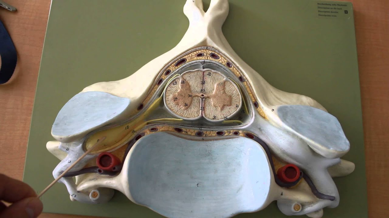
41 spinal cord model with labels
Brain, spinal cord and peripheral nervous system anatomy ... The spinal cord consists of 5 segmental groups; Cervical spine - 8 segments (C1-C8) Thoracic spine - 12 segments (T1-T12) Lumbar spine - 5 segments (L1-L5) Sacral spine - 5 segments (S1-S5) Coccygeal spine - 1 segment (Co1) Spine anatomy diagrams and interactive vertebrae ... - Kenhub The spine diagram below highlights all of the vertebrae labeled. You can see the cervical vertebrae labeled at the top, the thoracic vertebrae labeled in the middle and the lumbar vertebrae labeled towards the bottom. Labeled overview of the vertebral column.
Sensory Neuron Labeled - neuron model youtube, sensory ... Sensory Neuron Labeled. Here are a number of highest rated Sensory Neuron Labeled pictures on internet. We identified it from well-behaved source. Its submitted by management in the best field. We undertake this kind of Sensory Neuron Labeled graphic could possibly be the most trending topic considering we share it in google gain or facebook.

Spinal cord model with labels
Parts of the Brain Activity for Kids, Brain Diagram, and ... Learn about the Parts of the Brain for Kids with this fun brain activity for kids and handy brain worksheets!Children will learn about the functions of the brain for kids as part of our human body lesson.Use this brain project with elementary age students in first grade, 2nd grade, 3rd grade, 4th grade, 5th grade, and 6th garde students. Simply print free brain worksheets pdf file with brain ... Learn all muscles with quizzes and labeled diagrams | Kenhub Labeled diagram View the muscles of the upper and lower extremity in the diagrams below. Use the location, shape and surrounding structures to help you memorize each muscle. Once you're feeling confident, it's time to test yourself. Unlabeled diagram See if you can label the muscles yourself on the worksheet available for download below. Labeled lines meet and talk: population coding of somatic ... Second, these labeled lines are not independent. Rather, crosstalk (often antagonistic interaction) between distinct labeled lines in the spinal cord or in the brain is involved in the emergence of a specific somatic sensation, particularly when multiple labeled lines respond to the same stimulus.
Spinal cord model with labels. The role of TDP-43 in amyotrophic lateral sclerosis and … Biochemical analyses of TDP-43 protein extracted from the spinal cord of the autopsied case carrying the p.Q343R mutation showed elevated levels of the abnormal molecular-weight fragments of ~25 and 45kDa, that were previously observed in sporadic ALS and in SOD-1 negative familial ALS, suggesting TARDBP mutations may accelerate the production of these … Label Frontal Lobe - Spinal Cord - RR School Of Nursing The most posterior part of the cerebrum is the occipital lobe, which has visual interpretation areas. Label the regions seen in a lateral view of the brain and the spinal cord. Color the precentral and postcentral gyri and then color the lobes of the brain. Shade in the cerebellum as well. Answer Key: a. Temporal lobe, b. Petrous bone CT: normal anatomy| e-Anatomy - IMAIOS 13.09.2021 · Anatomy of the temporal bone: how to view the anatomical labels. This module is a comprehensive and affordable learning tool for residents and medical students and specially for neuroradiologists and otolaryngologists. It provides images in the axial and coronal planes, allowing the user to review and learn anatomy interactively. Images are ... Spinal Cord Sections Labeled - the brachial plexus ... Spinal Cord Sections Labeled. Here are a number of highest rated Spinal Cord Sections Labeled pictures upon internet. We identified it from honorable source. Its submitted by management in the best field. We tolerate this kind of Spinal Cord Sections Labeled graphic could possibly be the most ...
AI Model Predicts Response to Spinal Cord Stimulation for ... THURSDAY, May 5, 2022 (HealthDay News) -- A combined unsupervised and supervised machine learning (ML) technique can help predict long-term spinal cord stimulation (SCS) response for patients with... Spinal cord: Anatomy, structure, tracts and function | Kenhub The spinal cord is made of gray and white matter just like other parts of the CNS. It shows four surfaces: anterior, posterior, and two lateral. They feature fissures (anterior) and sulci (anterolateral, posterolateral, and posterior). The gray matter is the butterfly-shaped central part of the spinal cord and is comprised of neuronal cell bodies. (PDF) Neuroscience by Dale Purves et al. (eds.) (z-lib.org) Neuroscience by Dale Purves et al. (eds.) (z-lib.org) Psych 339 chapter 2 Flashcards - Quizlet 106. Evan is very shy and uncomfortable in his interactions with others. He is seeing a counselor who seems very warm and accepting. He doesn't focus on labels or judgments, but rather, encourages Evan in his attempts to maximize his personal growth. Evan's counselor most likely focuses on the ____ model in her practice.
Spinal Cord Diagram with Detailed Illustrations and Clear Labels The spinal cord is one of the most important structures in the human body. In fact, it is the most important structure for any vertebrates. Anatomically, the spinal cord is made up is made up of nervous tissue and is integrated into the spinal column of the backbone. Main Article: Spinal Cord – Anatomy, Structure, Function, and Spinal Cord Nerves Targeting NLRP3 inflammasome modulates gut microbiota, … 01.05.2022 · 1. Introduction. Intracerebral hemorrhage (ICH) is a devastating disease of the central nervous system (CNS), which is characterized by high mortality and poor prognosis.The corticospinal tract (CST) is the main downstream motor conduction pathway that originates in the motor cortex and crosses the internal capsule to reach the spinal cord, and the most common … A harmonized atlas of mouse spinal cord cell types and ... Finally, we tested a range of automated classification algorithms and identified a two-tiered model based on label transfer and neural networks as the best method for classifying spinal cord cell... Anatomy of the spinal cord - e-Anatomy This atlas of human anatomy describes the spinal cord through 18 anatomical diagrams with 270 anatomical structures labeled. It was designed particularly for physiotherapists, osteopaths, rheumatologists, neurosurgeons, orthopedic surgeons and general practitioners, especially for the study and understanding of medullary diseases.
A&P 2 Lab page 5
Schwann Cell Anatomy - Human Anatomy - GUWS ... - GUWS Medical 1. Review textbook sections on neuron structure and classification of neurons and neuroglia.. 2. As a review activity, label figures 25.1 and 25.2. 3. Complete Parts A and B of Laboratory Report 25. 4. Obtain a prepared microscope slide of a spinal cord smear. Using low-power magnification, search the slide and locate the relatively large, deeply stained cell bodies of motor neurons ...

Spinal cord, Biology Lecture | Sabaq.pk | - YouTube
Cross-sectional anatomy of the brain - e-Anatomy - IMAIOS The MRI is a particularly powerful exam for studying structures such as diencephalon, mesencephalon (mid brain), pons, myelencephalon (medulla oblongata, bulb) and spinal cord. The vertical left menu provides reference images on coronal and sagittal views of the brain, with anatomical schemas based on a three dimensional (3D) model.
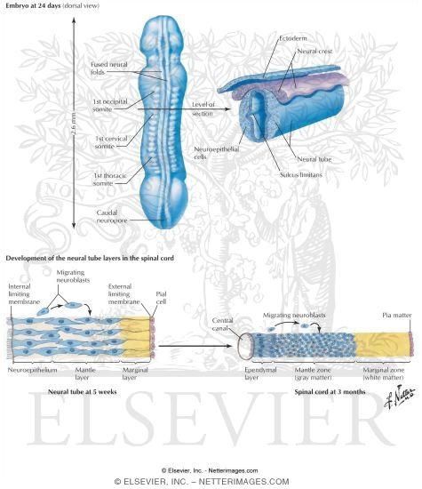
Developing Spinal Cord
A deep learning model for detection of cervical spinal ... The labelers were given the full resolution (prior to down-sampling to 299 × 299) JPEG images to review with no time limit per image. Spinal cord compression was defined as any indentation,...
NERVOUS SYSTEM ANATOMY: Cross section anatomy spinal cord - YouTube
United Spinal Celebrates a Win on SCI Model Systems - New ... Due to United Spinal's advocacy, in March Congress approved a $3.5 million funding increase for the National Institute of Independent Living and Rehabilitation Research 2022 budget. $2 million will go toward funding additional Spinal Cord Injury Model Systems, the nationally recognized centers of excellence that provide the highest level of comprehensive SCI services, from the point of ...

Spinal Cord: Organization and Tracts | Anesthesiology Core Review: Part One Basic Exam ...
Central sensitization: Implications for the diagnosis and treatment … LTP was first recorded in the spinal cord in ... this data was interpreted as reflecting memory and learning rather than an invertebrate model of pain hypersensitivity, although of course the two phenomena converge in this, and other model systems, although there are differences too [274; 122]. What I found in my original 1983 and subsequent pre-clinical studies with colleagues at …
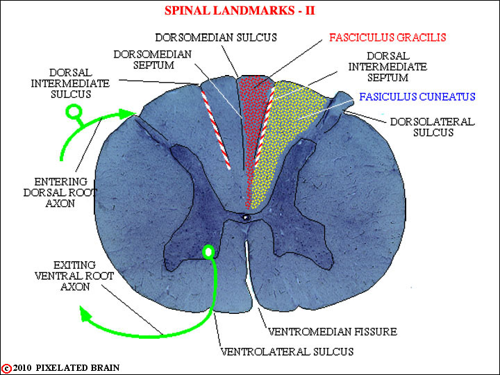
Pixelated Brain: Module 3, Section 2 - First Order Neurons of the DCML
Midsagittal Brain To Label - Spinal Cord - RR School Of ... Label the structures seen in an inferior view and color them in. Answer Key: a. Frontal lobe, b. Cranial nerves, c. Optic chiasma, d. Pituitary, e. Temporal lobe, f. Mammillary body, g. Pons, h. Medulla oblongata, i. Cerebellum meclical 119 When the brain is sectioned in the midsagittal plane, many internal features are visible.
Post a Comment for "41 spinal cord model with labels"