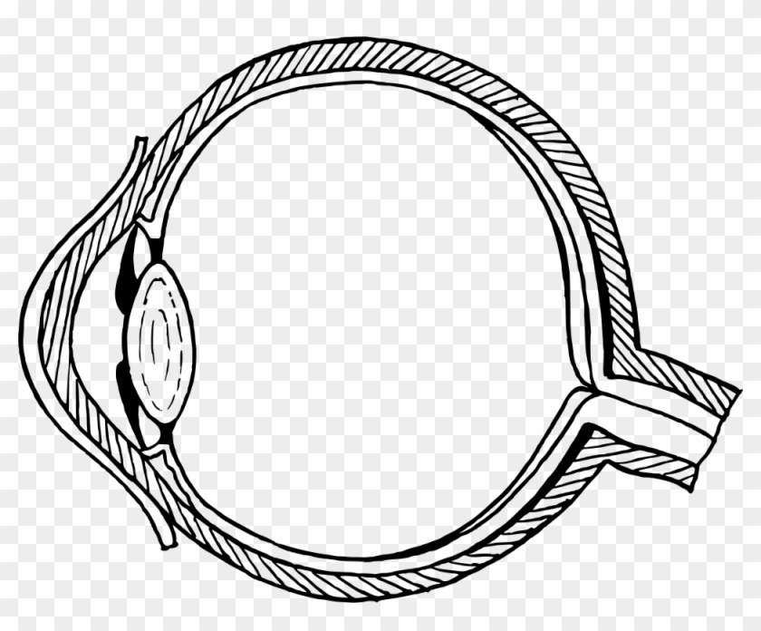
40 the human eye without labels
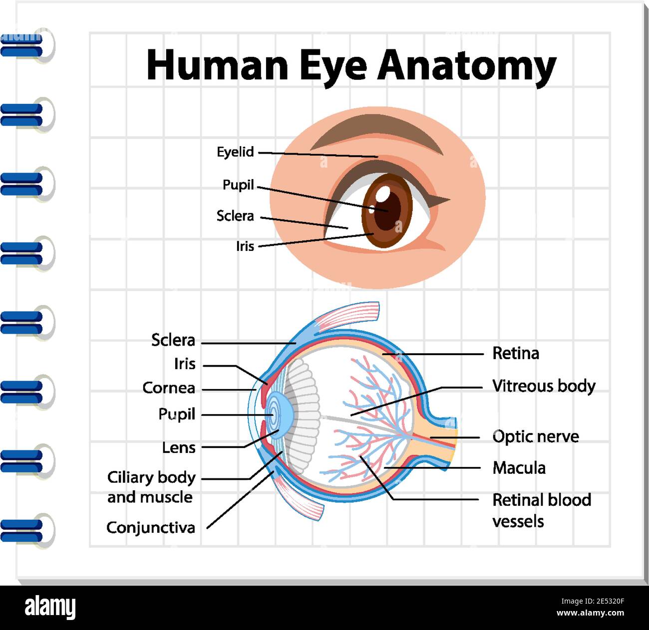
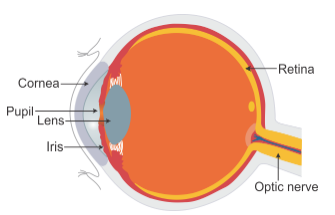
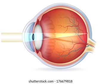
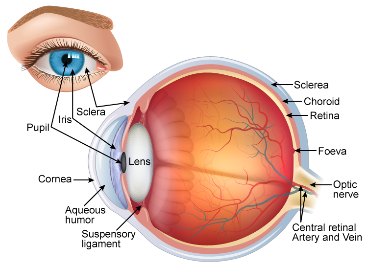
Structure and Function of the Human Eye - ThoughtCo The main parts of the human eye are the cornea, iris, pupil, aqueous humor, lens, vitreous humor, retina, and optic nerve. Light enters the eye by passing through the transparent cornea and aqueous humor. The iris controls the size of the pupil, which is the opening that allows light to enter the lens. Light is focused by the lens and goes ... What Does the Eye Look Like? - Diagram of the Eye | Harvard Eye Associates Vitreous Gel: A thick, transparent liquid that fills the center of the eye. It is mostly water and gives the eye its form and shape. Our eyes are vital for seeing the world around us. Keep them healthy by maintaining regular vision exams. Contact Harvard Eye Associates at 949-951-2020 or harvardeye.com to schedule an appointment today.
20 Facts About the Amazing Eye - Discovery Eye Foundation Here are a few facts you may enjoy: 1. Eyes began to develop 550 million years ago. The simplest eyes were patches of photoreceptor protein in single-celled animals. 2. Your eyes start to develop two weeks after you are conceived. 3. The entire length of all the eyelashes shed by a human in their life is over 98 feet with each eye lash having a ...

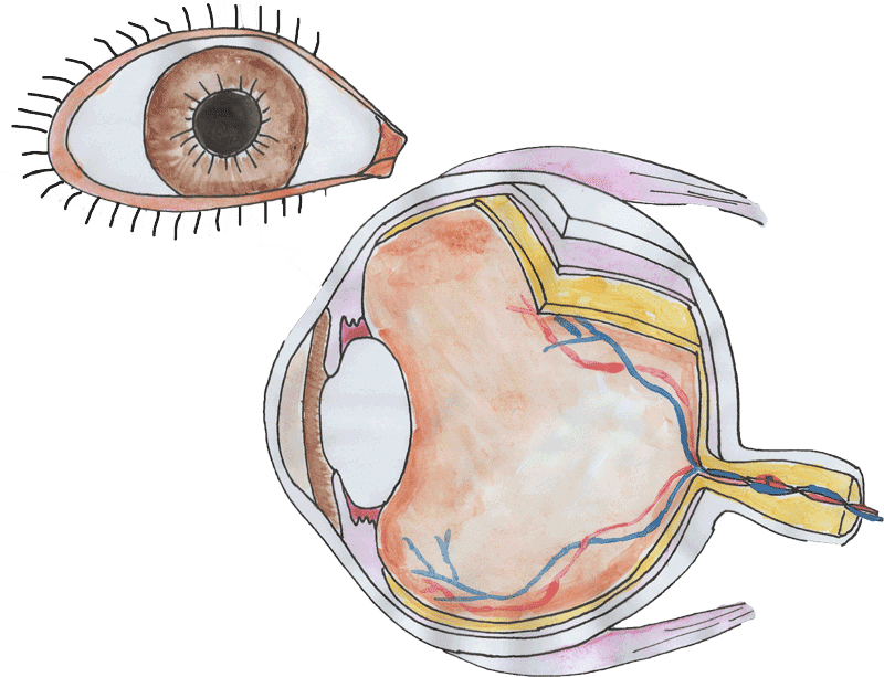
The human eye without labels
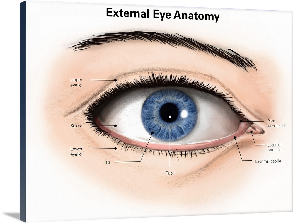
Eye Anatomy: A Closer Look At the Parts of the Eye - All About Vision In a number of ways, the human eye works much like a digital camera: Light is focused primarily by the cornea — the clear front surface of the eye, which acts like a camera lens. The iris of the eye functions like the diaphragm of a camera, controlling the amount of light reaching the back of the eye by automatically adjusting the size of the ... label the eye worksheet Eye From Front : Anatomy : The Eyes Have It. 9 Pics about Eye From Front : Anatomy : The Eyes Have It : Label the eye - ESL worksheet by zarawamp, Label School Supplies Worksheet — db-excel.com and also Eye From Front : Anatomy : The Eyes Have It. Eye From Front : Anatomy : The Eyes Have It kellogg.umich.edu. eye external anatomy lacrimal ... label muscles worksheet Picture Front Of The Eye Without Labels Clipart - Clipground clipground.com. eye human diagram worksheet layers eyeball learning anatomy labels without clipart eyes parts worksheets science structure clipground grade body structures. Posterior View Of The Superficial Muscles Of The Arm | ClipArt ETC etc.usf.edu
The human eye without labels. How Do We See Light? | Ask A Biologist - Arizona State University The human eye has over 100 million rod cells. Cones require a lot more light and they are used to see color. We have three types of cones: blue, green, and red. The human eye only has about 6 million cones. Many of these are packed into the fovea, a small pit in the back of the eye that helps with the sharpness or detail of images. How the Eyes Work | National Eye Institute - National Institutes of Health How the Eyes Work. All the different parts of your eyes work together to help you see. First, light passes through the cornea (the clear front layer of the eye). The cornea is shaped like a dome and bends light to help the eye focus. Some of this light enters the eye through an opening called the pupil (PYOO-pul). Eye Diagram With Labels and detailed description - BYJUS Iris is the coloured part of the eye and controls the amount of light entering the eye by regulating the size of the pupil. The lens is located just behind the iris. Its function is to focus the light on the retina. The optic nerve transmits electrical signals from the retina to the brain. Pupil is the opening at the centre of the iris. Label Parts of the Human Ear - University of Dayton Parts of the Ear. Select the correct label for each part of the ear. Click on the Score button to see how you did. Incorrect answers will be marked in red.
human eye | Definition, Anatomy, Diagram, Function, & Facts The protrusion of the eyeballs—proptosis—in exophthalmic goitre is caused by the collection of fluid in the orbital fatty tissue. The eyelids eyelid It is vitally important that the front surface of the eyeball, the cornea, remain moist. Human eye - Wikipedia The human eye is a sensory organ, part of the sensory nervous system, that reacts to visible light and allows us to use visual information for various purposes including seeing things, keeping our balance, and maintaining circadian rhythm . The eye can be considered as a living optical device. The Human Eye - Diagram, Parts, Working, Function and Work of The Lens Sclera: The sclera is the protective outer layer, a strong white coating that protects the eyes (white part of the eye). Cornea: The cornea is the sclera's translucent front part. The cornea allows light to flow through and into the eye. Iris: The iris is a black muscular tissue and ring-like structure behind the cornea. The eye's colour is determined by the colour of the iris. Label Parts of the Human Eye - University of Dayton Parts of the Eye. Select the correct label for each part of the eye. The image is taken from above the left eye. Click on the Score button to see how you did. Incorrect answers will be marked in red. ...
Anatomy and Structure of the Human Eye (With Diagrams) The iris is a flat, thin, ring-shaped structure sticking into the anterior chamber. This is the part that identifies a person's eye colour. The iris contains both circular muscles going around the pupil and radial muscles radiating toward the pupil. When the circular muscles contract, they make the pupil smaller. human eye diagram with labels Eye With Labels Clip Art at Clker.com - vector clip art online, royalty. 10 Images about Eye With Labels Clip Art at Clker.com - vector clip art online, royalty : Muscles of the Human Eyeball | ClipArt ETC, 35 Label The Structure Of The Eye - Labels Database 2020 and also 35 Label The Structure Of The Eye - Labels Database 2020. The Human Eye | Boundless Physics | | Course Hero The human eye is the gateway to one of our five senses. The human eye is an organ that reacts with light. It allows light perception, color vision and depth perception. A normal human eye can see about 10 million different colors! There are many parts of a human eye, and that is what we are going to cover in this atom. Properties label the ear worksheet Picture Front Of The Eye Without Labels Clipart 20 Free Cliparts clipground.com eye human diagram worksheet eyeball learning layers without anatomy labels parts eyes worksheets clipart science structure grade clipground body structures 14 Best Images Of Ear Hearing Worksheets - Listening Ear Craft Template
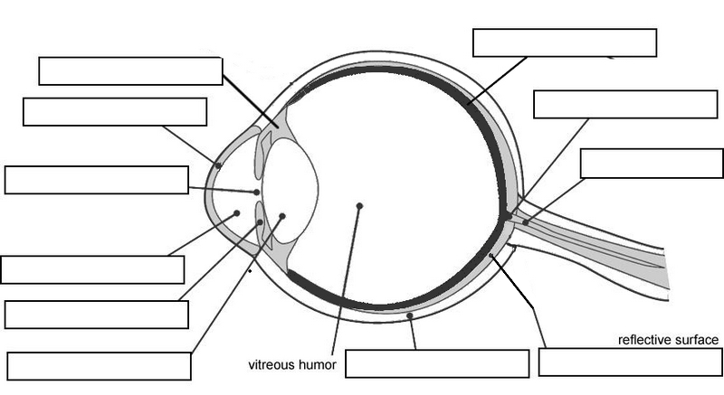
Anatomy of the eye: Quizzes and diagrams | Kenhub Here you can see all of the main structures in this area. Spend some time reviewing the name and location of each one, then try to label the eye yourself - without peeking! - using the eye diagram (blank) below. Unlabeled diagram of the eye Click below to download our free unlabeled diagram of the eye.
Human Eye Explorer Even microscopic structures, usually not visible for the human eye, can be explored: see inside the retina or cornea and discover its layers and cells from all perspectives. Additional functions like the presentation editor, the media library or labeling and color tools, make the Human Eye Explorer a unique software.
Quiz: Label The Parts Of The Eye - ProProfs Quiz Take up this quiz and find out how much did you get to understand about the human eye? All the very best to you! Questions and Answers 1. A is pointing to what part of the eye? A. Cornea B. Optic Nerve C. Iris D. Pupil E. Sclera 2. B is pointing to what part of the eye? A. Optic Nerve B. Lens C. Retina D. Pupil E. Iris 3.
Eye Anatomy: Parts of the Eye and How We See Here is a tour of the eye starting from the outside, going in through the front and working to the back. Eye Anatomy: Parts of the Eye Outside the Eyeball. The eye sits in a protective bony socket called the orbit. Six extraocular muscles in the orbit are attached to the eye. These muscles move the eye up and down, side to side, and rotate the eye.
File:Diagram of human eye without labels.svg - Wikimedia Size of this PNG preview of this SVG file: 410 × 430 pixels. Other resolutions: 229 × 240 pixels | 458 × 480 pixels | 732 × 768 pixels | 976 × 1,024 pixels | 1,953 × 2,048 pixels. Original file (SVG file, nominally 410 × 430 pixels, file size: 277 KB) File information. Structured data.
Eye Anatomy: 16 Parts of the Eye & Their Functions - Vision Center The lens of the eye (or crystalline lens) is the transparent lentil-shaped structure inside your eye. This is the natural lens. It is located behind the iris and to the front of the vitreous humor (vitreous body). The vitreous humor is a clear, colorless, gelatinous mass that fills the gap between the lens and the retina in the eye.
60,892 Human eye anatomy Images, Stock Photos & Vectors - Shutterstock Find Human eye anatomy stock images in HD and millions of other royalty-free stock photos, illustrations and vectors in the Shutterstock collection. Thousands of new, high-quality pictures added every day.
Category:Human eyes - Wikimedia Commons Black eyes by megamoto85 (cropped).jpg 925 × 673; 148 KB Blue Eyed Girl - Flickr - rcstanley.jpg 529 × 622; 49 KB Blue-Green Eye miosis.jpg 917 × 688; 451 KB
Structure of the Human Eye - Health Jade The eye is a hollow, spherical structure about 2.5 centimeters in diameter. Its wall has three distinct layers—an outer (fibrous) layer, a middle (vascular) layer, and an inner (nervous) layer. The spaces within the eye are filled with fluids that help maintain its shape. Figure 6. Structure of the human eye.
Human Eye - Definition, Structure, Function, Parts, Diagram - BYJUS Structure of Human Eye. A human eye is roughly 2.3 cm in diameter and is almost a spherical ball filled with some fluid. It consists of the following parts: Sclera: It is the outer covering, a protective tough white layer called the sclera (white part of the eye). Cornea: The front transparent part of the sclera is called the cornea.
The Eyes (Human Anatomy): Diagram, Optic Nerve, Iris, Cornea ... - WebMD The weaker eye, which may or may not wander, is called the "lazy eye." Astigmatism: A problem with the curve of your cornea. If you have it, your eye can't focus light onto the retina the way it...
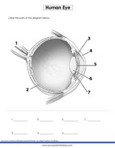
label muscles worksheet Picture Front Of The Eye Without Labels Clipart - Clipground clipground.com. eye human diagram worksheet layers eyeball learning anatomy labels without clipart eyes parts worksheets science structure clipground grade body structures. Posterior View Of The Superficial Muscles Of The Arm | ClipArt ETC etc.usf.edu
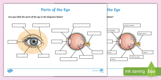
label the eye worksheet Eye From Front : Anatomy : The Eyes Have It. 9 Pics about Eye From Front : Anatomy : The Eyes Have It : Label the eye - ESL worksheet by zarawamp, Label School Supplies Worksheet — db-excel.com and also Eye From Front : Anatomy : The Eyes Have It. Eye From Front : Anatomy : The Eyes Have It kellogg.umich.edu. eye external anatomy lacrimal ...
Eye Anatomy: A Closer Look At the Parts of the Eye - All About Vision In a number of ways, the human eye works much like a digital camera: Light is focused primarily by the cornea — the clear front surface of the eye, which acts like a camera lens. The iris of the eye functions like the diaphragm of a camera, controlling the amount of light reaching the back of the eye by automatically adjusting the size of the ...















Post a Comment for "40 the human eye without labels"